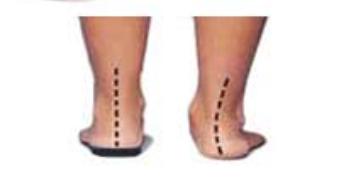
Description:
o Progressive flattening of medial longitudinal arch
o Was not present at birth (congenital flatfoot)
o Gait abnormality – excessive hindfoot valgus with initial ground contact
o Abduction of forefoot
o Foot fatigue from inadequate support of longitudinal arch
Above: From left to right, cavus deformity, normal foot, pes planus (flatfoot) deformity
Etiology:
o Posterior tibial tendon dysfunction
o Arthrosis (fusion) of tarsometatarsal (TMT) joints
o Charcot changes in midfoot due to peripheral neuropathy
o Talonavicular collapse from rheumatoid arthritis or trauma
o Usually no history of trauma
o Possible history of Lisfranc injury
Symptoms & Signs:
o Abducted forefoot
o Tiptoe standing – calcaneus remains in valgus, instead of normal inversion
o Too many toes sign
o Decreased or no active inversion strength
o Swollen, tender posterior tibial tendon
o Prominent medial cuneiform
o Charcot foot has variable swelling and deformity
o Rhematoid arthritis foot has a fixed valgus deformity of the hindfoot
Imaging:
o Radiographs usually elucidate cause of flatfoot deformity:
o Posterior tibial tendon dysfunction – sagging talonavicular joint or abduction of navicular on head of talus
o TMT arthrosis – joint degeneration and lateral and dorsal subluxation
o Charcot foot – dramatic joint destruction and dislocation
Rheumatoid foot – typical destructive, erosive changes with loss of bony architecture
Treatment:
Conservative
Support the longitudinal arch and ankle
Ankle-foot orthosis (AFO)
Must be shaped to accommodate any bony prominences to avoid skin breakdown
Surgical treatment
Depends on specific cause of flatfoot deformity:
Posterior tibial tendon dysfunction = reconstruction of tendon with flexor digitorum longus tendon
Possible calcaneal osteotomy if significant valgus deformity


