The Growth Plate
The growth plate is divided into multiple zones (Figure 8). These zones include the reserve zone, proliferative zone, and the hypertrophic zone. The reserve zone is the area closest to the secondary center of ossification. It has epiphyseal vessels the pass through this area but do not provide it with oxygen and keeps the oxygen tension low in this area. It has no known function with respect to longitudinal growth.
Figure 8
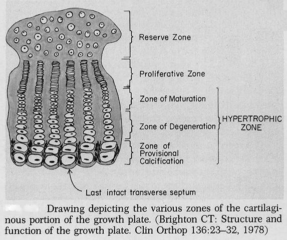
The proliferative zone contains a progenitor cell at the top of each flattened column of cells that is not derived from a cell within the reserve zone. These cells have large amounts of glycogen storage and highly active endoplasmic reticulum for protein synthesis. This region is responsive to hormones and mechanical factors. This region is affected in achondroplasia and a single mutation in the FGFR3 receptor will cause proliferative zone not to divide and subsequent failure of longitudinal growth.
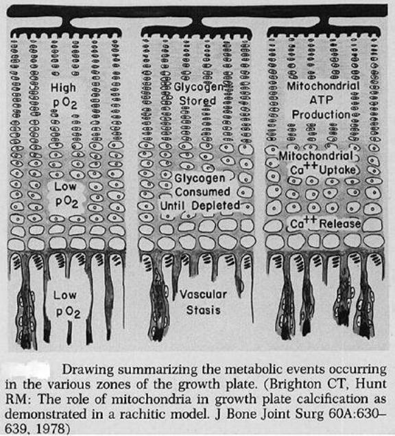
The hypertrophic zone is divided into three regions including: the zone of maturation, the zone of degeneration and the zone of provisional calcification. Throughout these zones the large quantities of glycogen stored in the proliferative zone are consumed while the cells transition from a high oxygen tension to low oxygen tension. The role of mitochondria change from one of energy production to calcium storage and eventual release. (Figure 9)
Figure 9
The nutrient artery that pierces the central region of the bone bifurcates and branches to form multiple metaphyseal arteries at the ends of the growth plate. These arteries have capillaries that perform a hairpin loop back upon themselves to provide venous return for this low-flow system. These vessels approach but do not penetrate the hypertrophic zone.
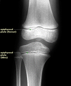
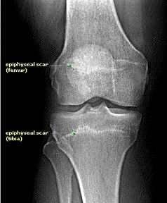
Blood vessels penetrate the epiphyses and form secondary centers of ossification through a similar process. When growth is complete, the progenitor cell stops dividing and the cartilage is entirely replaced by bone leaving only an epiphyseal “scar” (Figure 10).
Figure 10:
Open epiphyses top picture with closed epiphyses below leaving a “scar”.