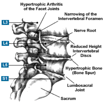
Description:
Any developmental of acquired narrowing of the spinal canal, nerve root canals, or intervertebral foramina that results in compression of neural elements
Pathophysiology:
- A small amount of canal narrowing occurs with age
- The narrowest part of the canal is L2 – L4
- Volume increases in flexion and decreases in extension
- Causes of pathologic narrowing of the canal include:
- Bulging of intervertebral discs posteriorly
- Buckling of the ligamentum flavum posteriorly
- Encroachment of the articular facets
- Degenerative spondylolisthesis
- Mechanical compression of cord results in increasing pressure in stenotic canal and:
- “neuroischemia” – nerve fibers are nutritionally deprived by compression of small vessels
- Inflammation of dura and exiting nerve roots results in adhesive arachnoiditis of the pia and friction neuritis which constricts and tethers neural elements
- Reduced permeability of hypertrophic restricts CSF flow, which provides 50% of nutrition to nerve fibers
- Pain and paresthesias are produced when activity increases the metabolic demands of nerve fibers beyond what the limited delivery of nutrients and removal of noxious substances allows
Classification:
- Congenital
- Example, achondroplasia
- Acquired – more common
- Degenerative
- Olisthetic-scoliotic
- Post-traumatic
- Post-operative
- Location of stenosis
- Central – hypertrophied structures put circumferential pressure around spinal cord
- Lateral – associated with narrowing of the foraminal canal

Signs/Symptoms of Degenerative Spinal Stenosis:
- Most common in elderly
- Women > Men
- Lower lumbar segments
- Insidious progression of lower back, buttock, thigh pain
- Lower extremity pain that is altered by position, relieved by rest – especially a position of flexion of the waist
- Can ambulate longer pushing a shopping cart (flexed body position)
- Distal pulses should be evaluated to distinguish claudication from neuroclaudication
Imaging:
- Plain radiographs demonstrate
- degenerative disc disease
- osteoarthritis of facets
- spondylolisthesis
- narrowing of interpedicular distance on AP
- CT scan allows for accurate measurement of canal dimensions
- Dural sac with diameter of less than 10mm correlates with clinical findings of stenosis
- MRI is now imaging modality of choice, comparable to contrast enhanced CT
Differential Diagnosis:
- Causes of referred pain to lower back:
- Retroperitoneal tumors
- Aortic aneurysms
- Peptic ulcer disease
- Renal lesions
- Hip and pelvis pathology
- Psychologic causes of low back pain
- Depression – common in elderly
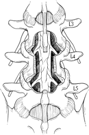
Typical midline decompression for spinal stenosis

Treatment:
- Non-steroidal anti-inflammatory medications
- Exercise program
- Many patients have appreciable response to NSAIDS and exercise
- Narcotics should be avoided
- Epidural corticosteroid injections have short-term success rate of 50% and long-term 25%
- Decompressive laminectomy has short-term success rate between 71-85%
- Reoperation common due to instability or recurrent stenosis
- Disc should be preserved for stability
- Prophylactic instrumented fusion should be performed if decompression will involve bilateral facet resection

