

Teambone has two research limbs:
The Basic Research limb was headquartered at the Bone and Joint Research Laboratory at the Veterans Affairs Medical Center in Salt Lake City, Utah (lab directors: Dustin Williams, Ph.D. and Roy Bloebaum, Ph.D.). This limb concentrates on bone’s adaptation to mechanical stimuli in the context of bone loss and osteoporosis. We are also working extensively to broaden the understanding of the bone organ from on microstructural, material and cellular level. We work closely with Dr. Kent Bachus, Director of the Bioengineering Research Laboratory.
The Applied Research limb is based at the Dr. Skedros’ clinic (Utah Orthopaedic Specialists) where Dr. Skedros has his clinical practice. The applied limb involves direct patient contact; our work in basic science can be directly translated into enhancing the quality of our patients’ lives. We are studying the diagnosis and treatment of osteoporosis and osteopenia and the orthopaedic surgeon’s role in treating problems stemming from skeletal senescence (including proximal humerus and femoral neck fractures). We also study the use of corticosteroid injections in treating painful shoulder conditions.
Research Themes:
Georgie Project
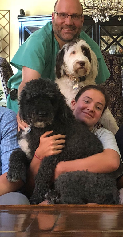
Dr. Skedros and his team are working on the Portuguese water dog “Georgie project” in collaboration with Dr. Gordon Lark (deceased) and Kevin Chase From the University of Utah Department of Biology.
Wolff Femoral Neck Work (ORS 2023-24)
Human Femur and Wolff’s Paradigm
Load Complexity Categories
Femoral Neck Stress Fractures and Wolff’s Paradigm
Basic Science Research Themes
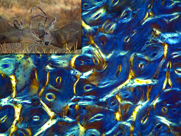 This is a circularly polarized light (CPL) image of a transversely sectioned mule deer antler from our recent study of antler histology (Skedros et al., 2014, J. of Structural Biology). This tissue was unstained and undecalcified, embedded in methyl-methacrylate, and diamond milled to 100 microns thick. Upper left: inset image of Rocky Mountain Mule deer fighting (photo courtesy of Robert McCleeary).
- Applications of backscattered electron imaging to bone mineralization and histomorphology
- Trajectorial theory of bone structure
- The role of collagen fiber orientation in bone adaptation
- The artiodactyl calcaneus as a model for bone examining cortical and cancellous bone adaptation
- Bone adaptation
- Structural and material organization of bones loaded in bending vs. torsion
- Advancing understanding of the load histories of proximal human and chimpanzee femora
Clincial Research Themes
OSTEOPOROSIS
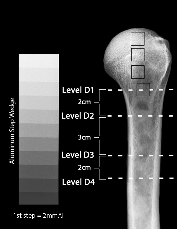
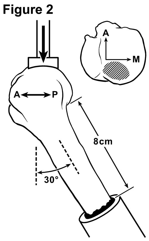 Figure 1 (at Left) shows an x-ray of a cadaver humerus bone (arm bone) next to an Aluminum step wedge. Figure 2 is a side view drawing showing a humerus being loaded in a way that simulates a backwards fall. In this study we are determining if x-ray measurements can be used to predict bone strength and to predict what kinds of methods would be best to repair the fracture (e.g., metal plates, pins, or artificial shoulder components).
- Factors that contribute to age-related increases in the rates of proximal humerus and femoral neck fractures.
- The role of the orthopaedic surgeon in diagnosing and treating osteoporosis
- The orthopaedic surgeon as a clinical densitometrist
- The coordination of discharge planning for patients with osteoporotic fractures (hospital admissions or E.R. visits)
SHOULDER AND ELBOW
- The use and misuse of corticosteroid injections in treating the painful shoulder conditions
- Advances in repair of rotator cuff tears (The “TOAKK” technique = Transosseous Anchor Double Knot)
- Evaluating radiographic density of the proximal humerus: is the bone normal or osteopenic?