

There are several deformities common to the hand that can occur as a result of traumatic injury, congenital abnormality, or degenerative disease states. Once hand deformities become relatively established, they can be difficult to significantly alter by splinting, exercise, or other nonoperative treatment.
Mallet Finger
Mallet finger is a flexion deformity of the terminal interphalangeal joint in which the fingertip droops and extension is not possible.
This deformity may result from a tendon injury or bony avulsion, causing a flexion lag at the distal interphalangeal (DIP) joint. Closed injuries may be treated with splinting that holds the DIP joint in extension and leaves the proximal interphalangeal (PIP) joint free. Avulsion fractures are usually united after 6 wk, but pure tendon injuries require approximately 8 to 10 wk for collagen to heal sufficiently.
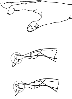
Swan-Neck Deformity
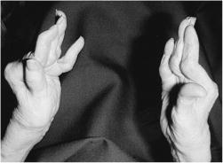
A swan neck deformity consists of metacarpal flexion, hyperextension of the PIP joint, and flexion of the DIP joint.
Although a characteristic finding in RA, swan-neck deformity has several causes, including untreated mallet deformity, volar plate laxity, spasticity, ligamentous laxity, and malunion of a fracture of the middle phalanx. The inability to overcome hyperextension of the PIP joint makes finger closure impossible and can cause severe disability. True swan-neck deformity does not affect the thumb, which is “missing” a phalangeal joint. However, severe hyperextension of the interphalangeal joint of the thumb with flexion of the metacarpophalangeal (MCP) joint is called a duck bill, Z (zigzag) type, or 90° angle deformity. If associated with thumb instability, swan-neck deformity can interfere greatly with prehension (pinch). This deformity can usually be corrected by interphalangeal arthrodesis.
Treatment is aimed at correcting the underlying cause when possible (ie, correcting the mallet deformity or any bony misalignment, rebalancing the extensor mechanism, releasing spastic intrinsic muscles).
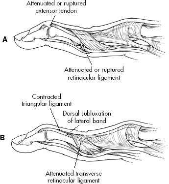
Boutonnière Deformity
(Buttonhole Deformity)
This is fixed flexion of the PIP joint accompanied by hyperextension of the DIP joint. It may result from lacerations, dislocation, fracture, osteoarthritis, or RA. Classically, the deformity is caused by disruption of the central slip attachment base of the middle phalanx extensor tendon, creating a so-called buttonholing of the proximal phalanx between the lateral bands of the extensor tendon. Surgical reconstruction after fixed deformities develop is often found to be unsatisfactory.

Erosive (Inflammatory) Osteoarthritis
A clinical form of osteoarthritis of genetic predisposition that, in the hand, primarily affects the DIP joint, some PIP joints, and the first carpometacarpal joints, with extensive synovitis and cyst formation.
Bony enlargement of the DIP joints (Heberden’s nodes) and bony overgrowth of the PIP joints (Bouchard’s nodules) are present, often without significant soft tissue swelling. The MCP joints and wrists are usually spared in erosive osteoarthritis. On x-ray, erosions appear subchondral rather than marginal (as is usually seen in RA). The thumb base (carpometacarpal joint) frequently is involved, with a squared-off appearance. Unlike in RA, the ESR and CBC are usually normal, regardless of disease severity. Treatment can include range-of-motion exercises in warm water, intermittent splinting to prevent deformity, use of analgesics or NSAIDs, and occasional intra-articular injections of depot corticosteroids for acutely symptomatic joints to relieve pain and prevent limited motion.
Congenital Hand Deformities
What are congenital hand deformities?
Congenital anomalies are deformities that are present at birth. Any type of deformity in a newborn infant can become a challenge for the child as he/she grows. Hand deformities can be particularly disabling as the child learns to interact with the environment through the use of his/her hands. The degree of deformity varies from a minor deformity, such as a digital disproportion, to a severe deformity, such as total absence of a bone.
Early consultation with a hand specialist is an important part of the treatment process for the child born with a hand deformity. Even if reconstructive surgery is not a possibility, there are many different types of prosthetic devices that can be used to increase function.
Classifications
The classifications for hand deformities can vary. The classifications listed below have been accepted by the American Society for Surgery of the Hand (ASSH). Classifications may change as more knowledge is obtained regarding each of the conditions. There are seven groups of deformities of the hand that will be discussed, including the following:
Problems in ontogeny (growth and development)
This occurs when parts of the body stop developing while the baby is in the womb, causing either a complete absence of a part of the body, such as the hand, or a missing structure, such as part of the arm bone. In the case of the complete missing part, surgery is not indicated. These children may be introduced to prosthetic devices early in their childhood. Types of this classification include, but are not limited to:
radial clubhand
A radial clubhand is a deformity that involves all of the tissues on the radial side (thumb side) of the forearm and hand. There may be shortening of the bone, a small thumb, or absence of the thumb. Deformities of the wrist are usually operated on around 6 months of age.
ulnar clubhand
An ulnar clubhand is less common than a radial clubhand. This deformity may involve underdevelopment of the ulnar bone (the bone in the forearm on the side of the little finger), or complete absence of the bone.
Failure of parts of the hand to separate
With this type of deformity, the parts of the hand, either the bones and/or the tissues, fail to separate in the womb. The most common type of this classification is syndactyly. Syndactyly is the most common congenital hand deformity, in which two or more fingers are fused together. According to some sources, this condition occurs in seven out of every 10,000 live births. There is a familial tendency to develop this deformity. This deformity usually involves both hands, and males are more often affected than females. If the fingers are completely fused together, it is considered complete. There are two types of syndactyly, including the following:
simple syndactyly – involves fusion between only the tissues of the fingers.
complex syndactyly – involves fusion between the bones.
Another example of failure of the hand to separate is seen in contractures of the hand. Contractures of the hand may also develop as a result of failure of the cells to differentiate in utero. A contracture is an abnormal pulling forward of the digits of the hand, usually caused by problems with the muscles or skin. One of the common types of this classification includes congenital triggering. Congenital triggering occurs when one of the digits is unable to extend. It is usually seen in the thumb. It may take some time in the child’s development before it is noted that the child can not extend the thumb. About 33 percent of these cases improve on their own. Surgery is usually not performed until the second year of life, but preferably before the age of 3.
Duplications of digits
Duplication of digits is also known as polydactyly. The little finger is the finger that is most often affected. There are three degrees of polydactyly, including the following:
type 1 polydactyly – an extra digit is attached by skin and nerves.
type 2 polydactyly – an extra digit with normal parts is attached to the bone or joint.
type 3 polydactyly – an extra digit with normal parts is connected to an extra normal metacarpal bone in the hand.
Undergrowth of digits
Underdeveloped fingers or thumbs are associated with many congenital hand deformities. Surgical treatment is not always required to correct these deformities. Underdeveloped fingers may include the following:
the digit is small
muscles are missing
bones are underdeveloped or missing
there is complete absence of a digit
Overgrowth of digits
Overgrowth of digits is also known a macrodactyly, which causes an abnormally large digit. In this situation, the hand and the forearm may also be involved. In this rare condition, all parts of the finger (or thumb) are affected, however, in most cases, only one digit is involved (usually the index finger). This condition is more commonly seen in males. Surgical treatment of this condition is complex and the outcomes may be less than desirable. Sometimes, amputation of the enlarged digit is recommended.
Congenital constriction band syndrome
This occurs when a tissue band forms around a digit (finger) or limb (arm), causing problems that can affect blood flow and normal growth. Ring constrictions are congenital (present at birth). This condition may be associated with other problems such as clubfoot, cleft lip, cleft palate, or other craniofacial anomalies. The cause of the ring constrictions is unknown. Some theories suggest that amniotic banding may lead to ring constrictions around a finger or limb.
With this condition, t here are four degrees of severity, including the following:
simple constrictions
constrictions that cause deformities beyond the constriction
constrictions that are associated with the fusion of parts of the finger
constrictions so severe that part of the finger will need to be amputated from the process
Treatment for congenital hand deformities:
Specific treatment for congenital hand deformities will be determined by your child’s physician based on:
the patient’s age, overall health, and medical history
extent of the condition
cause of the condition
the patient’s tolerance for specific medications, procedures, or therapies
expectations for the course of the condition
the physician’s opinion or preference
Treatment may include:
limb manipulation and stretching
splinting of the affected limbs
tendon transfers
external appliances (to help realign misshapen digits or hands)
physical therapy (to help increase the strength and function of the hand)
correction of contractures
skin grafts – involves replacing or attaching skin to a part of the hand that is missing skin or has been removed during a procedure.
prosthetics – may be used when surgery is not an option, or in addition to surgical correction.