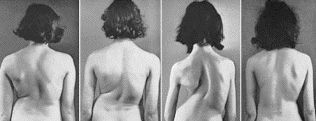
Four 70° curves, different patterns: Left-to-right lumbar, thoracolumbar, thoracic, and combined thoracic and lumbar curves.
Description:
- Curve is in coronal plane
- Often a rotational component
- Classified by age of onset – infantile, adolescent, adult
- Curvature named according to side of convexity and spinal level of the apex
- Incidence of 1% in general population
- Types of scoliosis:
- Congential scoliosis = curve 2° to structural bony abnormality
- Neuromuscular scoliosis = caused by neurologic disturbance or myopathy
- Idiopathic scoliosis = no determined cause, most common
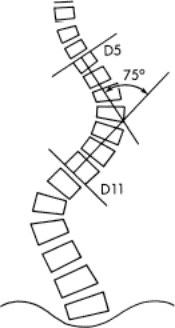
Cobb method for measuring curves.
- (1) locate the superior end vertebra and the
- (2) inferior end vertebra, and
- (3) draw intersecting perpendicular lines from the superior surface of the superior end vertebra and from the inferior surface of the inferior end vertebra. The angle of deviation of these perpendicular lines from a straight line is the angle of the curve. The end vertebra of the curve is the one that tilts the most into the concavity of the curve being measured.
Idiopathic Scoliosis:
- Right thoracic curve most common
- Right thoracic, left lumbar double curve second most common
- Compensatory curve = secondary curve to center head over pelvis
- Signs/Symptoms:
- Generally asymptomatic
- Asymmetry of shoulders, pelvis
- Rib hump, prominent scapula or lumbar fullness observed during “toe-touch”
- Curves of >60° result in cardiopulmonary compromise and restrictive lung disease
- Curve progression most common during periods of rapid skeletal growth: adolescence
- Radiographs:
- AP and lateral radiographs of entire spine, patient standing
- Curves measured using Cobb method (right)
- Flexibility or rigidity of curve can be determined by obtaining bending films
- Stagnara view for severe curves (>90°) = cassette parallel to rib hump, x-ray beam perpendicular cassette
Treatment of Idiopathic Scoliosis:
- Treatment is based on
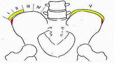
- Maturity of the patient (based on Risser’s stage (right)
- Presence of menarche
- Degree of deformity
- Curve progression
- Since most progression occurs at times of rapid growth, more frequent monitoring is required during adolescents
- Patients treated conservatively should be followed until the end of skeletal growth
- Girls grow until approximately 2 years after menarche
- Risser’s sign uses iliac apophysis to estimate remaining growth
- Hand and wrist physes can also be used to estimate remaining growth
Risser sign of skeletal maturity:
Stages represent presence of iliac apophysis (yellow) from not present (Stage 0 – skeletal immaturity) to completely fused (Stage V – skeletal maturity) Apophysis appears and fuses from lateral to medially.
Observation:
- Curves 10 or less require observation only
- Curves Adolescents with less than 2 years of growth remaining, no demonstrated progression of curve and a curve 40 are difficult to treat with bracing
- Severe curves at greater risk for progressing during adolescence and adulthood
- Many methods exist for surgical correction and stabilization of curves:
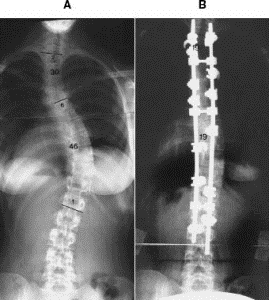
- Posterior fusion with Harrington rod instrumentation involves placing hooks on a ratcheted rod and distracting the ends, then performing fusion
- Variable hook-rod construct, such as the TSRH (Texas Scottish Rite Hospital) system.
- Multiple hooks along curve to apply distraction and compression at different levels


