

A. Fractures of the Tibial Plafond
Fractures of the tibial plafond are generally high-energy axial-loading injuries, such as in falls from heights or car wrecks. They are sometimes called pilon fractures after the French for “hammer” or “pestle,” referring to the manner in which the talus strikes the plafond. They are also occasionally seen in twisting injuries to the ankle, in which a large fracture of the medial or posterior malleolus extends to the weight-bearing surface of the joint. Theses injuries are seen most frequently in young men (ages 35-40), and are rare in children and the elderly.
Physical Exam –
In tibial plafond fractures, massive soft tissue swelling and compromise are the rule, consistent with a high-energy mechanism. Evaluation of the soft tissues, including inspection and probing of open wounds, noting the presence and character (serous vs hemorrhagic) of fracture blisters, and determining the degree of swelling by the presence of skin wrinkles is critical. A careful neurovascular exam both before and after reduction is important in fracture dislocations. Finally, these factors should be reassessed periodically, as the degree of soft tissue injury may not be apparent initially, and development of a compartment syndrome may occur as late as 24 hours following injury.
As high-energy fractures, these injuries are frequently associated with associated injuries (27-51%).
Radiographs and Classification –
AP, lateral, and mortise views should be obtained initially. Post-reduction films, traction films, and 45º oblique films of the ankle may provide additional information in evaluating the fracture. CT scans and films of the contralateral ankle may be useful sub-acutely for pre-operative planning.
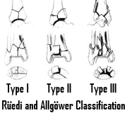
The Reüdi and Allgöwer system is frequently used to classify plafond injuries based on degree of plafond displacement and comminution. Type I is a non-displaced articular fracture, type II is displaced at the articular surface but not comminuted, and type III has significant comminution at the articular surface. The AO/OTA classification system is much more complicated, dividing the fractures into 27 subgroups based on the general classifications of extraarticular, partial articular, and total articular.
Treatment –
Initial treatment of pilon fractures involves relieving the ongoing compromise of the soft tissues. This requires reduction of the ankle joint, immobilization, and minimization of swelling with elevation and ice. Holding a reduction may require calcaneal traction (Böhler frame) or spanning external fixation acutely. Open wounds may require one or more irrigation and debridement prior to definitive fixation.
Operative treatment of pilon fractures may be considered early, before the soft tissues become excessively swollen (usually in the first 8-12 hours). Definitive treatment of pilon fractures are often delayed 7-10 days to allow soft tissues to recover from the initial insult. Open reduction and internal fixation is standard of care for most pilon fractures, providing anatomic alignment of the joint surface. External fixation, either spanning with calcaneal traction or on the same side of the joint with a skinny-wire fixator may be considered for definitive treatment as well.
Non-operative treatment in a long-leg cast may be appropriate for low-energy, non-displaced fractures or fractures in severely debilitated people.
Treatment of pilon fractures carries a high incidence of complications. Wound complications may occur in 30-40%. Malunion, non-union, shortening, and post-traumatic arthritis also occur frequently.
B. Ankle Fractures
Ankle fractures are a common fracture occurring in the elderly. Incidence in persons over age of 60 is 130/100,000 in some series. This fracture is most common in elderly women. Increased body weight and smoking are two risk factors for ankle fractures.
History and Physical Exam –
Ankle fractures are commonly seen with a twisting (low-energy) mechanism of injury. The position of the foot and the force acting on it are important factors in predicting the pattern of fracture and the likely treatment (based on the Lauge-Hansen classification). Screening for associated injuries is important because they occur frequently in association with ground-level falls in elderly, osteoporotic women.
The Ottowa Ankle Rules are a screening device used to determine who among adults presenting with ankle injury should receive radiographs. Age > 55, inability to bear weight, and bone tenderness at posterior edge or tip of either malleolus should lead to obtaining radiographs. These factors lead to a sensitivity of nearly 100% for detecting fracture in the setting of pain near the malleoli.
Ankle injuries frequently present with pain and swelling at the lateral malleolus. Tenderness around the medial malleolus is often an important determinant of stable versus unstable ankle fractures. Assessment of the syndesmosis may be aided by the squeeze test, in which the leg is squeezed 5cm proximal to the ankle joint and produces pain at the syndesmosis. Examination of the fibula by palpation for proximal tenderness will help recognize Maisonnueve fractures.
Anatomy –
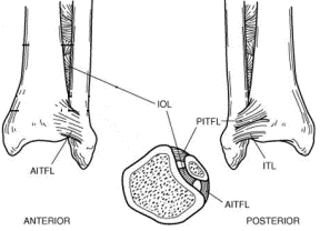
The ankle joint is composed of three bones and multiple ligaments that provide it with stability. The bony arch of the ankle mortise joint is composed of the medial mallelous off of the tibia, the lateral malleolus, which is composed of the distal extent of the fibula, and the tibial plafond, the weight-bearing surface of the distal tibia. The syndesmotic ligaments join the tibia and the fibula together distally and provide stability to the mortise as it overlies the saddle-shaped talus. The syndesmotic ligaments are the anterior inferior tibiofibular ligament (AITFL), the posterior inferior tibiofibular ligament (PITFL), the inferior transverse ligament (ITL). The interosseous ligament also provides stability to the mortise. A posterior malleolus off of the plafond provides anterior-posterior stability to the ankle joint.
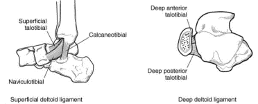
The talus is held beneath the ankle mortise by strong medial and weaker lateral ligaments. Medially, the deltoid ligament is made up of a superficial portion (composed of the calcaneotibial, talotibial, and naviculotibial ligaments, arising from the anterior colliculus of the medial malleolus) and a deep portion (composed of the deep anterior talotibial and deep posterior talotibial ligaments, arising from both the anterior and posterior colliculi). These deep deltoid ligaments serve as the primary stabilizers of the ankle. The lateral collateral ligaments of the ankle are the anterior and posterior talofibular ligaments and the calcaneofibular ligament. The anterior talofibular ligament is the weakest of the three, and is susceptible to injury in inversion ankle sprains.
Radiographs and Classification –
Basic radiographic evaluation of the ankle consists of three views – the lateral, the AP (in line with the second metatarsal), and the mortise view (internally rotated 15º). In addition to evaluating for fractures, several indicators of joint alignment should be evaluated. The medial clear space, from the medial malleolus to the medial talus, is an indicator of patency of the deltoid ligaments, the primary stabilizers of the ankle. It should be approximately equal to the space between the distal tibia and the talus on the mortise view (greater than 4mm is abnormal). The patency of the syndesmosis may be evaluated by evaluating the tibiofibular clear space (which should be 6mm or less) and the tibiofibular overlap (which should be 1 cm or greater) on the AP view. Weight-bearing or stress radiographs may be helpful in demonstrating subtle syndesmotic injury or ankle instability.
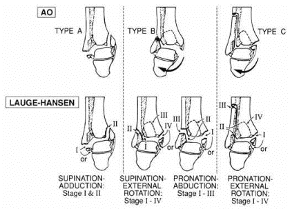
The Lauge-Hansen classification of ankle fractures defines each injury by the position of the foot (supination or pronation) and the deforming force acting on it (external rotation, abduction, adduction). A predictable pattern of injury results from the various combinations: supination-adduction (SAD), supination-external rotation (SER), pronation-abduction, and pronation-external rotation (PER). This classification system is useful conceptually and in the rare occasion when a displaced ankle fracture is treated closed (reversing the injuring force reduces the fracture).
The Weber classification is based on the level of the fibula fracture in an ankle injury. This classification is simple and clinically useful. Weber A fractures are fractures of the fibula below the level of the syndesmosis and generally represent stable ankle fractures. Weber B fractures are fractures of the fibula at the level of the syndesmosis (usually the syndesmosis remains intact), and represent unstable fractures only if associated with medial injury (medial malleolar fracture or deltoid ligament disruption). Weber C fractures are fibula fractures above the syndesmosis and generally represent unstable fractures with syndesmotic disruption.
Treatment –
Treatment of ankle fractures is based on the stability of the ankle joint. Stable ankle fractures are those in which the talus remains centered under the tibia, meaning that no medial-sided injury (with resultant lateral shift of the talus) has occurred. Some examples of stable fractures include most Weber A fractures and Weber B fractures without medial injury. Stable fractures may be treated non-operatively with protected weight-bearing (below-knee cast or walking boot).
In unstable fractures, the talus is not held centered under the tibia by the mortise joint; this may be due to loss of bony stability (Weber B fibula fractures with medial malleolar fracture or deltoid ligament disruption) or ligamentous stability (syndesmotic injuries with widening of the mortise associated with medial injury). Unstable ankle fractures will demonstrate lateral shift of the talus (with widening of the medial clear space) on plain radiographs or stress views. Unstable ankle fractures are generally treated by ORIF. In bimalleolar injuries (medial and lateral malleoli fractures), this generally involves plating of the fibula, along with fixation of the medial malleolus, using either screw fixation or tension-band techniques. In injuries with ligamentous medial injury, usually fixation of the medial malleolus fracture will restore ankle stability. Syndesmosis injuries require ORIF, usually one or two screws from the fibula into the tibia. Rarely, unstable fractures may be treated by closed reduction and immobilization in a long-leg cast.