The movements of the hand are accomplished by two sets of muscles and tendons: the flexors, for bending the fingers and thumb, and the extensors, for straightening out the digits. The flexor muscles are located on the anterior (volar) surface of the forearm and are attached by tendons to the phalanges of the fingers. The extensor muscles are on the dorsal surface of the forearm and are similarly connected. The thumb has two separate flexor muscles that move the thumb in opposition and make grasping possible.
The Skeleton of the Hand
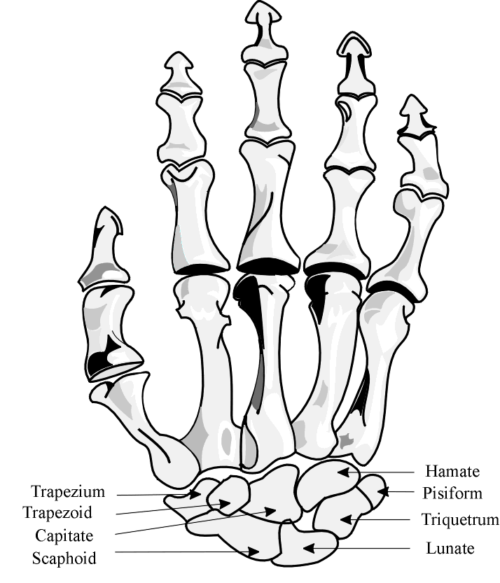
The hand has 27 bones: the 8 bones of the carpus (wrist), arranged in two sets of four; the 5 bones of the metacarpus, one to each digit; and the 14 digital bones, or phalanges, 2 in the thumb and 3 in each finger. The carpal bones fit into a shallow socket formed by the bones of the forearm. Each metacarpal bone has a proximal base, a shaft, and a distal head. The base of each metacarpal contacts the distal row of carpal bones to form thecarpometacarpal joint. The head of each metacarpal contacts the proximal
at a metacarpophalangeal (MP) joint.
The proximal row of phalanges articulates with the middle row of phalanges at the proximal interphalangeal (PIP) joints, the middle and distal rows of phalanges articulate at the distal interphalangeal (DIP) joints.
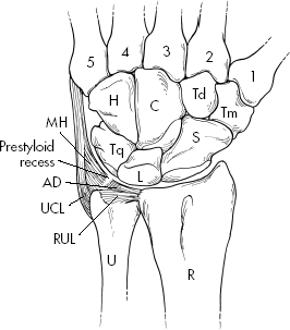
The carpal bones can be remembered using the mnemonic “Some Lovers Try Positions That They Can’t Handle”
Proximal Row (Radial to ulnar wrist)
Distal Row (Radial to ulnar wrist) Trapezium/Greater multangular (That)
|
| Muscles of the hand | |
|
|
-
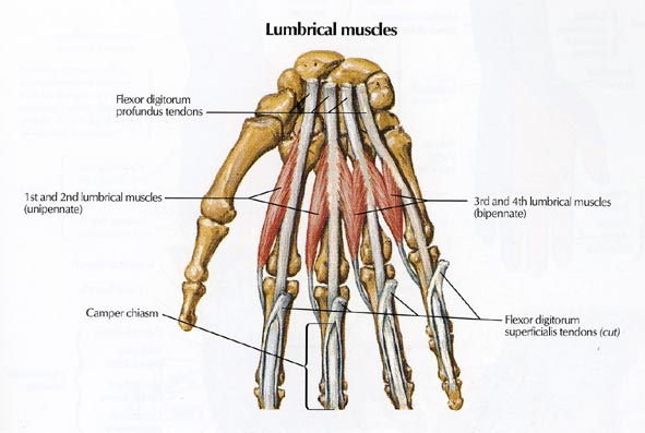
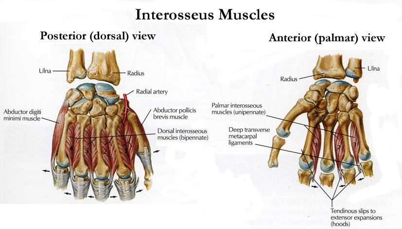
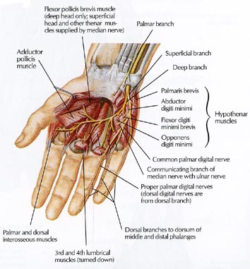
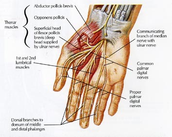
Intrinsic Muscles of the Hand Muscle Origin Insertion Action Innervation Muscles acting on the Second through Fifth Digits Dorsal interossei (4) ulnar side of 1st MC; both sides of 2nd-4th MC; radial side of 5th MC Tubercle of proximal phalanx and dorsal aponeurosis: radially on 2nd and 3rd digits, ulnarly on 3rd and 4th digit Abduct 2nd-4th digits from midline; flex MCP joint, extend PIP and DIP joints Ulnar nerve (C8-T1, anterior) Palmar interossei (3) ulnar side of 2nd and radial side of 4th-5th MC Tubercle of proximal phalanx and dorsal aponeurosis: ulnarly on 2nd digit, radially on 4th and 5th digits Adduct 2nd, 4th, and 5th digits to the midline of the hand. Flex MCP joint and extend PIP and DIP joints Ulnar nerve (C8-T1, anterior) Lumbricals (1 and 2) Tendons of FDP in deep palm radial side of dorsal expansion of 2nd and 3rd digits Flex MCP, extend DIP, PIP Median nerve (C8-T1, anterior) Lumbricals (3 and 4) Tendons of FDP in deep palm radial side of dorsal expansion of 4th and 5th digits Flex MCP, extend DIP, PIP Ulnar nerve (c8-T1, anterior) Palmaris brevis ulnar border of palmar aponeurosis Skin over hypothenar region Corrugates palmar skin Ulnar nerve (C8-T1, anterior) Abductor digiti minimi Pisiform bone Ulnar side of base of 5th proximal phalanx Abduct 5th digit Ulnar nerve (C8-T1, anterior) Flexor digiti minimi brevis Flexor retinaculum and hamulus Ulnar side of base of 5th proximal phalanx Flex 5th MCP joint Ulnar nerve (C8-T1, anterior) Opponens digiti minimi Flexor retinaculum and hamulus Ulnar side of base of 5th MC Flexion and opposition Ulnar nerve (C8-T1, anterior) Muscles acting on the Thumb Abductor pollicis brevis (APB) Anterior surface of trapezium, scaphoid radial aspect of base of proximal phalanx Abducts thumb Median n. (C8-T1, anterior) Opponens pollicis Trapezium Anterolateral surface of 1st MC Medially rotates (opposes) thumb Median n. (C8-T1, anterior) Flexor pollicis brevis Superficial head Transverse carpal ligament and trapezium radial side of base of proximal phalanx Flexes thumb Median n. (C8-T1, anterior) Deep head radial side of 2nd MC Ulnar nerve (C8-T1, anterior) Adductor pollicis Oblique head Anterior surface of capitate and 2nd and 3rd MC ulnar side of base of proximal phalanx Adducts thumb Ulnar nerve (C8-T1, anterior) Transverse head Distal half of 3rd MC - Extrinsic (Originate in forearm)The extrinsic muscles of the hand include the wrist flexors and extensors, which stabilize the wrist in slight dorsiflexion, the Finger Flexors (five deep and four superficial), and the finger extensors
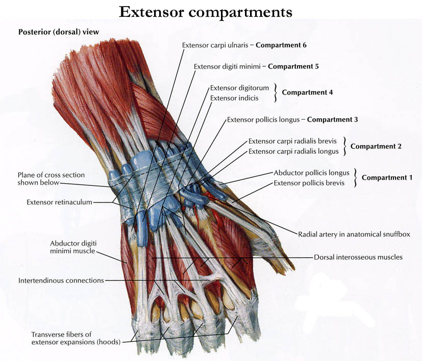
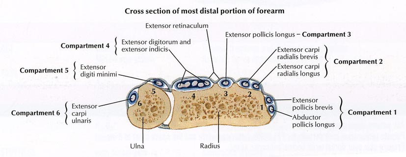
| Extrinsic Muscles of the Hand | ||||
| Muscle | Origin | Insertion | Action | Innervation |
| Muscles acting on the Second through Fifth Digits | ||||
| Extensor digitorum cominus (EDC) | Lateral epicondyle of humerus | middle and distal phalanges of index, middle and ring fingers | Extends digits and wrist when fist is clenched | Radial nerve (C7-C8, posterior) |
| Extensor digiti minimi (EDM) | Common extensor tendon | All phalanges of fifth digit | Extends fifth digit | Radial nerve (C7-C8, posterior) |
| Extensor indicis proprius (EIP) | Interosseus membrane and ulna | middle and distal phalanges of index finger | extends first digit and wrist | Radial nerve (C8-T1, posterior) |
| Flexor digitorum superficialis (FDS) | Medial epicondyle | Base of middle phalanx of each digit | Flexes PIP, MCP, and wrist joint | Median nerve (C8-T1, anterior) |
| Flexor digitorum profundus (FDP) | Anterior proximal ulna and IOM | Base of distal phalanx of each digit | Flexes DIP, PIP, MCP, and wrist joints | -Median n. (C7-T1, anterior) for 2nd-3rd digit -Ulnar n. (C7-T1, anterior) for 4th-5th |
| Muscles acting on the thumb | ||||
| Abductor pollicis longus (APL) | Posterior IOM and ulna | Base of1st MC, laterally | Abducts thumb and wrist | Radial n. (C8-T1, posterior) |
| Extensor pollicis brevis (EPB) | Posterior midshaft of radius and IOM | Base of proximal phalanx | Extends thumb and abducts wrist | Radial n. (C8-T1, posterior) |
| Extensor pollicis Longus (EPL) | Posterior surface of IOM and posteriior ulna | Base of middle phalanx | Extend thumb and abducts wrist | Radial n. (C8-T1, posterior) |
| Flexor pollicis longus (FPL) | Anterior mid-radius and IOM | Lateral aspect of base of proximal phalanx | Flexe thumb, MCP joint, and wrist | Median n. (C7-T1, anterior) |
| Blood Supply | |
| The blood supply to the hand is via the ulnar and radial arteries. The ULNAR artery continues to the hand at the medial (ulnar) side of the wrist.The deep palmar branch arises just distal to the lateral side of the pisiform bone, where the ulnar pulse is generally palpable. This branch then passes through the hypothenar compartment to the deep palmar arch by anastamosing with the terminal branch of the radial artery.The superficial palmar branch is the major contributor to the superficial palmar arch, which lies just beneath the palmar aponeurosis. This arch is larger than the deep arch. The common digital arteries arise from this arch; each bifurcates into proper digital branches, which run on the medial and lateral side of adjacent digits in a neurovascular bundles, containing digital artery, vein, and nerve.The RADIAL artery continues to the hand at the lateral (radial side of the wrist. The superficial palmar branch of the radial artery arises at the level of the end of the radius and branches just before the radial artery passes through the “anatomic snuff box”, which is bounded by the EPL and EPB tendons and is where a radial pulse may be palpated. The superficial palmar branch then passes through the thenar compartment, and provides a minor contribution to thesuperficial palmar arch. The deep palmar branch of the radial artery is the major contributor to the deep palmar arch. It courses around the base of the thumb and passes dorsal to the 1st MC to gain access to the thenar space where it becomes the deep palmar arch. |
|

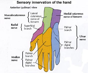
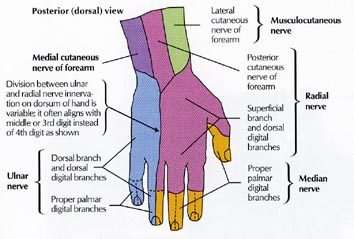
| Innervation to the Hand | |
| The hand is innervated by three main nerves, the ulnar, medial and radial nerves. | |
The ULNAR nerve enters the hand with the ulnar artery
|
|
The MEDIAN Nerve enters the hand through the carpal tunnel
|
|
The RADIAL Nerve courses with the radial artery.
|
|
Wrist structures
|