

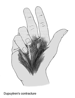
Many people find that as they age it becomes increasingly difficult to fully extend or stretch their fingers. In many cases, arthritis or some other joint disease may be to blame. But a condition in the palm of the hand may also cause the fingers to contract. Dupuytren’s (pronounced du-pwe-trahns’) contracture is a fairly common condition that occurs when the connective tissue (fascia) under the skin begins to thicken and shorten. As the tissue tightens, it may pull the fingers down towards the palm of the hand. In some individuals, the condition may progress until the involved fingers become disabled.
Dupuytren’s disease is a proliferative fibroplasia of the subcutaneous palmar tissue, occurring in the form of nodules and cords, that may result in secondary progressive and irreversible flexion contractures of the finger joints. Other secondary changes include thinning of the overlying subcutaneous fat, adhesion to skin, and later pitting or dimpling of the skin.
The activity of the lesion and the rate of deformity are variable. Occasionally a finger may become markedly flexed within a few weeks or months, but the development of a severe deformity usually requires several years. In some patients the lesion progresses steadily, whereas in others exacerbations and remissions occur; however, regression is rare.
Approximately 5% of patients with Dupuytren contractures have similar lesions in the medial plantar fascia of one or both feet, known as Ledderhose disease, and 3% of patients demonstrate plastic induration of the penis, known as Peyronie disease. “Knuckle pads” are common on the dorsum of the proximal interphalangeal (PIP) joints. Patients with these associated findings are considered to have a Dupuytren diathesis and are prone to progressive and recurrent disease.
Commonly occurring in the fifth to seventh decades of life, Dupuytren contracture occurs 10 times more frequently in men than in women. According to McFarlane the disease occurs significantly earlier in men (33 to 63 years) than in women (46 to 70 years). It is most common in those of Scandinavian and Celtic origin, although it occasionally has been reported in blacks and rarely in Asians. The lesion has been reported to be more frequent and severe in persons with diabetes mellitus or with epilepsy (42%), and conflicting reports exist concerning the disease in persons suffering from alcoholism. The involvement, although often bilateral (45%), rarely is symmetrical. Mikkelsen et al. found that mortality may be increased in men who develop the disease before the age of 60.
Etiology
Although the causes of this disease are unknown, trauma to the hand and the type of manual labor performed by an individual may be contributing factors. The presence of hemosiderin in these lesions suggests hemorrhage from tears; however, the nondominant hand is as often affected as the dominant one, thus making trauma alone an unlikely cause. Some believe that occasionally a single injury may precipitate the onset of the disease in genetically susceptible individuals. According to McFarlane, a causal relationship may be assumed if consistent histological changes occur within 2 years after a focal injury in women younger than 50 years of age and men younger than 40 years of age without a strong diathesis for Dupuytren disease. In patients with bilateral disease, the disease should develop in the uninjured hand after the age of 40 in men and 50 in women.
Evidence also points to heredity as a predisposing factor in Dupuytren disease. The lesion seems to occur earlier and more frequently in some families, and James has suggested an autosomal dominant pattern. Vascular insufficiency and cigarette smoking have been linked to Dupuytren disease as possible causative factors.
The lesion usually begins in line with the ring finger at the distal palmar crease and progresses to involve the ring and little fingers, these digits being affected more frequently than all others combined. Flexion contractures of the metacarpophalangeal and PIP joints gradually develop, their severity depending on the extent and maturity of the fibroplasia. Discomfort is rare and usually consists of itching or occasional pain early in the development of the nodules.
Pathogenesis
In 1972 Gabbiani and Majno implicated the myofibroblast as the dominant cell type in Dupuytren contracture. This is a contractile cell with increased type 3 collagen, possibly originating from a transformed perivascular smooth muscle cell. The Dupuytren fibroblast also has higher levels of alpha smooth muscle actin and contractility than a normal fibroblast. Most investigators now agree that in Dupuytren contracture the subcutaneous nodules and cords are formed by fibroplasia and by hypertrophy of already existing fibers of the palmar fascia. Millesi believes that the pathological tissue arises only through changes in the existing fibers of the palmar fascia and not by the formation of new tissue. Luck suggested that the subcutaneous nodules develop first and cords form proximally in response to intermittent stress resulting in contractures. According to this concept the nodules progress through proliferative, involutional, and residual stages. In the proliferative stage, the nodules are young, non-stress aligned fibroblasts that expand and displace the subcutaneous tissues and fuse to the skin. The nodules typically appear about the distal palmar crease over the metacarpophalangeal joint and distally over the PIP joint, but never over the distal interphalangeal joint. The nodules eventually stop growing and begin to contract in the involutional stage. Stress alignment of the fibroblasts occurs and more collagen is produced. Nodule contraction places tension on the normal fascia proximally, producing fascial hypertrophy and nodule-cord units. In the residual phase, the nodules decrease in size and may become acellular fibrous cords. Gosset believes that the nodules and cords do not represent two stages of the disease but rather two forms originating in two different tissues, the subcutaneous fat and palmar fascia. Hueston believes that the nodules develop subcutaneously and only later may involve the palmar fascia and overlying skin. Nevertheless, contractures of the metacarpophalangeal and PIP joints, as well as displacement of digital neurovascular bundles, result from predictable patterns of fascial cord involvement.
TABLE 1 — Terminology
Normal (band)
Diseased (cord)
Pretendinous band
Pretendinous cord
Vertical septa of Legeau and Juvara
Not affected
Central fibrofatty tissue
Central cord
Deep extension of Gosset
Spiral cord
Superficial transverse ligament
Not affected
Natatory ligament of Grapow
Natatory cord
Lateral digital sheet of Gosset
Lateral cord
Grayson ligament and lateral cords
Parts of central, spiral, and lateral cords
Cleland ligament
Parts of spiral and lateral cords
Landsmeer ligament
Not affected
From McFarlane RM: Plast Reconstr Surg 74:31, 1974.
The fascial structures that may become involved in the fibroproliferative process have been clearly outlined by McFarlane (Table 1). Various authors have paid considerable attention to tissue terminology. The longitudinally oriented fascia located dorsal to the neurovascular bundle, termed the retrovascular cord is involved in the disease and may be implicated as a cause for recurrent PIP contractures. The Cleland ligament generally is believed to be spared. The pretendinous cord nearly always is responsible for primary contracture of the metacarpophalangeal joint. It may attach to the distal palmar crease skin, base of the proximal phalanx, or the tendon sheath at this level, or it may extend to attach to the flexor tendon sheath over the middle phalanx or the skin in this area. A spiral cord occurs when four normally existing structures (pretendinous band, spiral band, lateral digital sheet, and the Grayson ligament) become diseased. The spiral cord runs dorsal to the neurovascular bundle proximally and volar to it distally. When the spiral cord is contracted, the neurovascular bundle is drawn toward the midline of the finger. Neurovascular displacement is found most commonly on the ulnar aspect of the little and ring fingers, and tedious dissection is required to prevent digital nerve injury.
The lateral digital cord may extend distally and contribute to a flexion contracture of the distal interphalangeal joint. The plane between this cord and the overlying skin is minimal and must be developed sharply. The retrovascular cord is not believed to contribute significantly to flexion contracture of the PIP joint; however, it may be responsible for some residual flexion contracture or recurrence if not excised.
Metacarpophalangeal and distal interphalangeal joint contractures appear to result from pretendinous and lateral cord development. However, PIP joint contractures may develop from isolated digital cords in addition to central, spiral, or retrovascular cords. According to Strickland and Bassett, the diseased tissue in this unusual form of Dupuytren contracture most commonly affects the small finger; however, any digit may be involved. The cord originates from the periosteum of the proximal phalanx and fascia overlying the intrinsic muscles and distally courses dorsal to the neurovascular bundles, inserting into the middle phalanx or the overlying flexor tendon sheath volar to the neurovascular bundles. This digital cord frequently displaces the neurovascular bundle superficially to the midline of the finger, similar to a spiral cord.
Signs and symptoms
The first sign is a thickening (nodule) in the palm of the hand that most frequently develops near the base of the ring or little finger. The nodule, which can resemble a callus, is painless but may be tender to the touch. Gradually, other nodules may develop and extend a contracture across the first joint into the finger. The overlying skin begins to pucker, and rough cords of tissue extend into the finger. As the process continues, these cords tighten and pull the finger in toward the palm. The ring finger is usually affected first, followed by the little, long and index fingers. The problem is not pain, but the restriction of motion and the deformity it causes.
The progress of the disease is often sporadic and unpredictable. Exactly what triggers the formation of nodules and cords is unknown. As the disease progresses, the diseased tissue wraps itself around and between normal tissue.
Many people do not seek medical care until the contracture is well advanced. The only treatment for this condition is surgery, which is usually reserved for individuals who have developed deformity as a result of the progressive contracture. Because many nodules do not progress to contracture and because scar tissue from previous surgeries can make excision of recurrent nodules more difficult, surgical removal of isolated nodules is not indicated in most cases.
A good guideline for determining when to consider surgery is the “table top test.” Try to place the palm of your hand completely flat on a hard surface. If you can’t, the contracture has progressed to a point where surgical intervention could be helpful.
Prognosis
The prognosis in Dupuytren contracture seems to depend on the following factors, which in turn may determine the extent of any operation.
Heredity. A family history of the disease is an indication that the lesion is likely to progress more rapidly than usual, especially if the onset is early.
Sex. The lesion usually begins later and progresses more slowly in women, who often accommodate better to the resulting deformity. Zemel et al. have shown, however, that long-term results after operation are worse in women than in men, with postoperative flare reaction being twice as likely.
Alcoholism or epilepsy. The lesions are more severe, progress more rapidly, and recur more frequently when associated with these conditions.
Location and extent of disease. When the disease is bilateral and especially when it is associated with knuckle pads and nodules in the plantar fascia, progression is more rapid and recurrence more frequent. Progression is more rapid on the ulnar side of the hand.
Behavior of disease. How the disease has behaved in the past, whether treated or not, is an indication of its probable behavior in the future.
Treatment
Many medical remedies have been tried; the most common treatment is surgical. Reports on clinical trials evaluating clostridial collagenase injections as a nonoperative treatment have indicated prompt and impressive metacarpophalangeal and PIP joint contracture release; however, this treatment is still in clinical trials at this time. In the absence of contractures, treatment usually is not indicated because nodules and cords usually are painless. Rarely is the presence of a palmar nodule alone an indication for surgery unless sufficient discomfort, pitting, and maceration occur. Patients with slowly progressing, nondisabling contractures should be examined periodically, perhaps every 3 months. Surgical treatment is seemingly technically easier when joint contractures are smaller; however, many patients will have ill-defined planes between normal and abnormal tissue when the cellular process is active. Ideally, patients are operated on when their disease is more mature and the tendency for surgical trauma to accelerate the disease process is less. Nonetheless, PIP joint and metacarpophalangeal joint contractures of 15 and 30 degrees or more usually are disabling and may warrant surgical intervention. Stiffening and increase in flexion contractures may occur when surgical intervention occurs in the proliferative stage. Moreover, indications for and timing of surgery should also take into account the disability of the joint contracture(s), presence of degenerative joint disease, and other predisposing factors for poor outcomes rather than merely the degree of contracture.
Five common surgical procedures used in treating Dupuytren contracture are
(1) subcutaneous fasciotomy,
(2) partial (selective) fasciectomy,
(3) complete fasciectomy,
(4) fasciectomy with skin grafting, and
(5) amputation.
The appropriate procedure depends on the degree of contracture, nutritional status of the palmar skin, the presence or absence of bony deformities, and the patient’s age, occupation, and general health. In general, more severe involvement requires more extensive surgery carried out in stages if necessary and preceded perhaps by a subcutaneous fasciotomy and joint extension therapy.
Subcutaneus fasciotomy. The least extensive procedure, subcutaneous fasciotomy, commonly is used for elderly patients who are not concerned with the appearance of the disease or in patients who have poor general health. The results of this procedure are better in the residual phase when dense, mature cords are present than when the lesions are more immature and diffuse. Rodrigo et al. believe that fasciotomy should be considered a temporary measure, because 43% of contractures so treated in their study required repeat surgery.
Partial (selectiv) fasciectomy. Partial (selective) fasciectomy usually is indicated when only the ulnar one or two fingers are involved. This operation is used more frequently because postsurgical morbidity is less and complications are fewer than after complete fasciectomy. Even though the rate of recurrence after partial fasciectomy is 50%, the need for another surgical procedure is only 15%. In this operation only the mature deforming tissue is excised. However, it must be emphasized that all diseased tissue may not be removed, since biochemically or microscopically involved fascia may not be clinically apparent at the time of surgery (Albin et al., 1975). Various incisions can be used to expose the pathological tissue. Some prefer a zigzag incision on the fingers or a variant of it because exposure of the diseased tissue is better. However, the incision should be fashioned to fit the needs of the individual patient, considering the contractures and the adherence of skin to the underlying fascia. When tightness of the palmar skin limits extension of a finger, a midline incision converted to appropriate Z-plasties is indicated. Regardless of the incision, dissection is made easier by loupe magnification.
Sometimes after fasciectomy, extension of the PIP joint is incomplete. Projections of isolated cords passing volar to the rotation axis of the PIP joint are not uncommon causes of residual joint flexion deformity. These problematic cords often insert onto the flexor tendon sheath laterally or the middle phalanx. Dissection of faintly detectable deforming cords intimately associated with the skin, as well as skin Z-plasty, may be required for sufficient PIP joint contracture correction. However, significant residual PIP flexion contractures may require volar joint capsulotomies. PIP joint flexion contractures of more than 60 degrees are correctable to about 50% of the existing contracture, regardless of a concomitant PIP joint capsulotomy.
The technique of Skoog is a partial or selective fasciectomy in which only the pretendinous fibers of the palmar fascia are excised. According to Skoog, there is a definite plane between the pretendinous longitudinal fibers of the palmar fascia and the transverse palmar ligament that is limited to the midpalmar area. He emphasizes, however, that the pretendinous fibers may seem to be attached to the ligament. Further, according to Skoog, the interdigital or natatory ligaments do become involved in Dupuytren contracture and prevent the fingers from spreading normally and are distinguishable from the transverse palmar ligament by their more distal location.
Complete fasciectomy. Complete fasciectomy rarely if ever is indicated because it is associated frequently with complications of hematoma, joint stiffness, and delayed healing, and it does not completely prevent recurrence of the disease. Fasciectomy with skin grafting may be indicated for young people in whom the prognosis is poor because of such factors as epilepsy, alcoholism, the presence of the disease elsewhere in the body, and recurrence of the lesion after prior excision. The skin and underlying abnormal fascia are excised, and a full-thickness or split-thickness skin graft is applied. Recurrence has not been reported in areas of the palm treated in this manner.
Amputation. Amputation, although rarely necessary, may be indicated if flexion contracture of the PIP joint, especially of the little finger, is severe and cannot be corrected enough to make the finger useful. A 40-degree flexion contracture usually is tolerated fairly well. The skin from the involved finger can be used to cover a palmar skin defect; the finger is filleted and the skin is folded into the palm as a pedicle with its neurovascular bundles.
Another alternative for the severely contracted PIP joint is joint resection and arthrodesis. This results in a shortened finger but avoids the potential for recurrent PIP joint contracture and a potential amputation neuroma.