

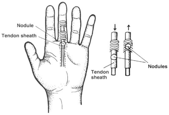
Trigger finger is an often painful snapping, triggering, or locking of the finger as it is flexed and extended. This is due to a localized inflammation or a nodular swelling of the flexor tendon sheath that doesn’t allow the tendon to normally glide back and forth under a pulley. It occurs in the superficial and deep flexor tendons adjacent to the A1 pulley at the metacarpal head (Figure below). The thumb and the middle and the ring fingers of the dominant hand of middle-aged females are most commonly affected. It is often encountered in patients with diabetes and rheumatoid arthritis. The relationship of trigger finger to repetitive trauma has been frequently cited in the literature; however, the exact mechanism of this correlation is still open for debate.
 |
Symptoms
Patients typically complain of pain in the proximal interphalangeal (PIP) joint of the finger, rather than in the true anatomic location of the problem—at the metacarpophalangeal (MCP) joint. Some individuals may report swelling and/or stiffness in the fingers, particularly in the morning. Patients may also have intermittent locking in flexion or extension of the digit, which is overcome with forceful voluntary effort or passive assistance. Multiple finger involvement can be seen in patients with rheumatoid arthritis or diabetes. |
Examination
The essential element in the physical examination is the localization of the disorder at the level of the MCP joint. There is palpable tenderness, and sometimes a tender nodule, over the volar aspect of the metacarpal head. Swelling of the finger may also be noted. Opening and closing of the hand actively produces a painful clicking as the inflamed tendon passes through a constricted sheath. Passive extension of the DIP or PIP joint while keeping the MCP joint flexed may be painless and done without triggering. With chronic triggering, patient may develop IP joint flexion contractures. Therefore it is important to determine whether there is normal passive range of motion in the MCP and interphalangeal (IP) joints. Neurologic examination including strength, sensation, and reflexes should be normal with the exception of severe cases that are associated with disuse weakness and/or atrophy. Co-morbidities can affect the neurologic exam as well (e.g., someone with diabetic neuropathy may have impaired sensation).
Functional Limitations
Functional limitations include difficulty with grasping and fine manipulation of objects due to pain, locking, or both. The patient may have problems with typing, buttoning a shirt, driving a car, using tools at work, etc.
Diagnostic Studies
This is a clinical diagnosis. Patients without a history of injury or inflammatory arthritis do not need routine radiographs. MRI can confirm tenosynovitis of the flexor sheath, but this offers minimal advantage over clinical diagnosis.
Differential Diagnosis
| Anomalous muscle belly in the palm |
| Dupuytren’s disease |
| Ganglion of the tendon sheath |
| Tumor of the tendon sheath |
| Rheumatoid arthritis |
Treatment
The initial goal of treatment is to restore the normal gliding of the tendon through the pulley system. This can often be achieved with conservative treatment. However, typically the first line of treatment is a local steroid injection. The determination whether to inject first or try non-invasive measures is often based on the severity of the patient’s symptoms (more severe symptoms generally respond better to injections), the level of activity of the patient (e.g., someone who needs to get back to work as quickly as possible) and the patient’s and clinician’s preferences.
Non-invasive measures generally involve splinting the MCP joint at 10 to 15 degrees for up to 2 weeks. This has been reported to be quite effective, although less so in the thumbs. Also, DIP splinting provides a reliable and functional means of treating work-related trigger finger without lost time from work. Additional conservative treatment includes icing the palm (20 minutes 2 to 3 times per day in the absence of vascular disease), NSAIDs/COX-2 inhibitors, and avoidance of exacerbating activities. Wearing padded gloves provides protection and may help to decrease inflammation by avoiding direct trauma.
Rehabilitation
Rehabilitation may include treatment with an occupational or physical therapist experienced in the treatment of hand problems. Supervised therapy is generally not necessary but may be useful in the following scenarios:
(1) when a patient has lost significant strength, range of motion, and/or function from either not using the hand, from prolonged splinting, or post-operatively;
(2) when modalities such as ultrasound or iontophoresis are recommended to reduce inflammation; and
(3) when a customized splint is deemed to be necessary.
Therapy should focus on increasing function and decreasing inflammation and pain. This can be done by utilizing techniques such as ice massage, contrast baths, ultrasound, and iontophoresis with local steroid use. For someone with a very large or small hand or other anatomic variations (e.g., arthritic joints), a custom splint may fit better and allow him or her to function at work more easily than a pre-fabricated splint. Improving range of motion and strength can be done via supervised therapy either prior to surgery or postoperatively.
| Procedures
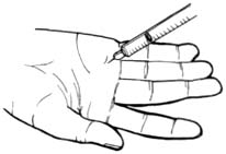
A local steroid injection (Figure right) can be used as an alternative or in addition to other management. The steroid injections are usually beneficial and frequently curative. However, they may need to be repeated up to 3 times. This procedure is less effective with multiple digits involvement (such as in patients with diabetes or rheumatoid arthritis) or when the condition has persisted greater than 4 months. |
|
Surgery
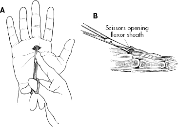
Although steroid injections should be tried for most trigger finger cases prior to considering surgery, surgical intervention is highly successful for conservative treatment failures and should be considered for patients desiring quick and definitive relief from this disability. Individuals with diabetes and rheumatoid arthritis are more likely to require surgery. There are two general types of surgery for this condition: the standard operative release of A1 pulley and the percutaneous A1 pulley release procedure. Both surgical procedures are generally effective and carry a relatively low risk of complications.
Potential Complications
Disease-related complications include permanent loss of range of motion from a contracture developing in the affected finger—most commonly at the PIP joint. In rare instances, chronic intractable pain may develop despite treatment.
Potential Treatment Complications from NSAIDs are well known (gastric, renal, and hepatic). COX-2 inhibitors may have fewer gastric-associated side effects. Complications from local steroid injections include skin depigmentation, skin atrophy, tendon rupture, digital sensory nerve injury, or infection. Individuals with rheumatoid arthritis are more likely to have tendon rupture; therefore, repeated injections are not recommended in these cases. Possible surgical complications include infection, nerve injury, and flexor tendon bowstringing.