Skip to content
Description:
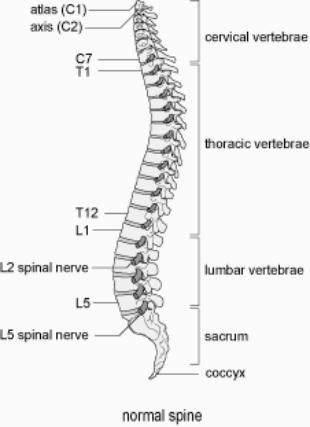
- The spinal column typically consist of 33 vertebrae. Seven cervical, 12 thoracic, and five lumbar vertebrae compose the movable presacral spine; five fused segments form the sacrum with four or five irregular ossicles caudal to the sacrum forming the coccyx
- The gross anatomic appearance is one of cervical lordosis, thoracic kyphosis, and lumbar lordosis
- The typical vertebrae consists of two major components: a roughly cylindrical ventral mass of cancellous bone, the body and the dorsal vertebral arch
- The arch is attached to the dorsolateral aspects of the body by two stout pillars, the pedicles. These are united dorsally by a pair of arched flat laminae that are surmounted in the midline by a dorsal projection, the spinous process.
- The pedicles, laminae, and dorsum of the body thus form the vertebral foramen, a complete osseous ring that encloses the spinal cord.
- Near the junction of the pedicles and the laminae are found the lateral transverse processes and the superior and inferior articular processes. The transverse processes extend from the sides of the vertebral arches. The articular processes form the facet or zygapophyseal joints.
- The pars interarticularis is the portion of the lamina that lies between the superior and inferior articular processes. It sees the dorso-ventral translational forces which has been implicated in pars fractures.
- An endplate composed of hyaline cartilage lies on both the superior and inferior endplates of the vertebral bodies. Between them lie the intervertebral disc composed of a circumferential annulus fibrosis and a central nucleus pulposus. The endplates are important in the diffusion of nutrition and waste between the blood supply of the vertebral bodies and the avascular discs.
Cervical Spine:
- In the cervical spine C1 and C2 have a distinct morphology compared to C3-C7.
- C1, the atlas, is a ring of bone that supports the base of the skull at the atlanto-occipital articulation. Half of the flexion and extension of the neck occur at this articulation.
- C2, known as the axis, provides a bearing surface on which the atlas may rotate, but its most distinctive characteristic is the vertically projecting odontoid process that serves as a pivotal restraint against horizontal displacements of the atlas.
- Crucial stabilizing ligaments maintain the relationships between the occipito-atlanto-axial articulations. Most notable is the transverse atlantal ligament which spans between the atlantal condyles trapping the odontoid anteriorly against the anterior arch of C1. This ligament keeps C1 from sliding forward on C2 during flexion. While approximately half of the rotation of the neck occurs at the C1-C2 articulation, it is limited by the alar ligaments spanning from the odontoid to the anterior rim of the foramen magnum.
- The remainder of cervical vertebrae are similar in morphology as described. The lateral edges of their vertebral bodies turn sharply upward to form the uncinate processes.
- This is only present in the cervical spine. The transverse processes in the cervical spine also contain vertebral artery foramina which transmit the vertebral arteries within them from C6 to C1, prior to the arteries penetrating the posterior atlanto-occipital membrane and joining to become the basilar artery.
- From the subaxial cervical spine inferiorly down to L5, the basic components of vertebral structure are similar.
Ligaments of the Spine:
- Supporting ligaments give the column stability. The anterior (ALL) and posterior longitudinal ligaments (PLL) run from the foramen magnum to the sacrum.
- The ALL largely inserts into the central aspect of the vertebral bodies.
- The PLL primarily attaches to the superior and inferior edges of the vertebral bodies and bowstrings across the central portion.
- The ligamentum flavum is a very strong ligament that runs within the canal attaching to the undersurface of the laminae. From segment to segment, the fibers of the ligamentum flavum insert into the superior edge of the inferior lamina and then blend with the undersurface of the superior lamina.
- An interspinous ligament attaches the stalks of the spinous process while a supraspinous ligament attaches to the tips of the spinous processes. Together, these two ligaments serve as a tension band and act as a tether when the spine is flexed. These ligaments provide the spinal column with flexible stability.
Nerve Anatomy:
- The neurologic component of the spine is often misunderstood. The naming of the nerve roots in relation to their exiting level changes from the cervical to the thoracic spine.
- Because there are seven cervical vertebrae and eight cervical nerves, the cervical nerve roots exit above the pedicle of their corresponding level. That is, the C1 nerve root exits above the C1 pedicle. At C7, the C7 nerve root exits above the C7 pedicle and the C8 nerve root exits below the C7 pedicle.
- This then sets up the pattern where the remainder of caudal nerve roots exit below their corresponding pedicle. It can, therefore, be expected that the L4 nerve root exits below the L4 pedicle. The understanding of this relationship is central to understanding the pathology of disc herniation.