Introduction
Bones have multiple functions that include structural support and protection for vital organs, support and leverage for movement and manipulation, repository for essential minerals and housing for the hematopoietic system.
Bone Composition
Thirty-five percent of bone is composed of a flexible extracellular matrix consisting mostly of collagen. The remaining sixty-five percent is composed of harder, more rigid materials. These mainly consist of calcium and phosphorous (mostly in the form of hydroxyapatite). The cellular components of bones include osteoprogenitor cells, osteoblasts, osteocytes, and osteoclasts (Figure 1). Osteoprogenitor cells are unspecialized bone cells with a mesenchymal origin that possess mitotic ability and can differentiate into osteoblasts. These cells are found throughout the bone with heavy concentrations around the endosteum and periosteum. Osteoblasts are cells that produce the extracellular substances of bone tissue. Osteoblasts lack mitotic capacity and are derived strictly from osteoprogenitor cells. Often a transitional cell called a pre-osteoblast can be seen on micrographs (Figure 2). They are found throughout the bone and concentrate on the surfaces of the bone where their activity is stimulated by calcitonin produced within the thyroid gland. Osteoblasts that become encased in extracellular substances are known as osteocytes. Osteocytes lose capacity for bone production and function in a bone maintenance role where they support the existing extracellular environment. Finally, osteoclasts are multinucleate cells that function to break down existing bone. They are usually found on bone surfaces and lead the tips of “cutting cones” (Figure 3), which are required to remodel existing bone. They are derived from monocytes and are positively regulated by parathyroid hormone.
Figure 1
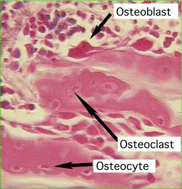
Figure 2:
Histologic slide of the different stages of osteoblast. Overlying these cells is the periosteum.
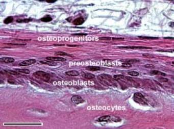
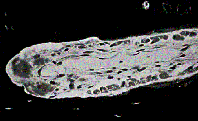
Figure 3:
Below is pictured a cutting cone with an osteoclast at it’s leading edge (left). See the section on remodeling for more details.
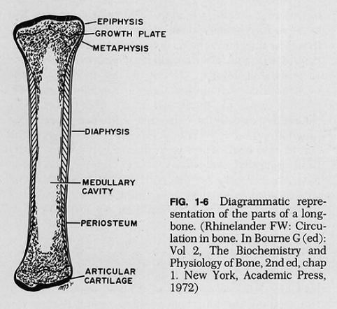
There are two basic forms of bone. The first is called woven bone (also known as primary bone), which describes an immature, disordered bone that is often formed rapidly. This bone is found in the early stages of fracture healing and is the primary component of early callus (Figure 4). It has a low mineral content and disordered composition. Woven bone will remodel to a mature form of bone called lamellar bone. There is a highly ordered structure to the lamellar bone, called an osteon. Within a bone there are two configurations for osteons that include the cortical and cancellous regions. These configurations are distributed in variable amounts thru the three regions and are called the epiphysis, metaphysis and diaphysis (Figure 5). High-density cortical regions are found within the metaphysis and diaphysis of bones and are typically arranged into hollow cylinders that are adapted to resisting bending forces. Within the metaphysis, the cortical regions begin to thin out and the lower density cancellous regions predominate. These regions are particularly adapted to supporting weight bearing by flaring out to provide a larger surface area and increasing porosity to help resist strain. This helps distribute the forces of impact to the bulk of the bony structure during activities such as walking or running. The epiphysis describes the most distal aspect of the bone that contains the articular cartilage and subchondral bone.
Figure 4:
Note the callus formation thru the healed 3rd metatarsal fracture.
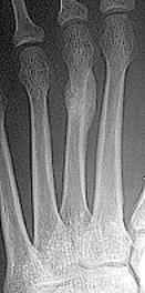
Figure 5:
Idealized bone anatomy