Bone Formation
There are two types of bone formation that occur during development. The first is called intramembranous ossification and is the primary method of forming the clavicles and skull bones. In this process, osteoblasts cluster within fibrous membranes to form a center of ossification. They begin secreting collagenous fibers that are subsequently calcified until trabeculae are formed. Eventually the osteoblasts are surrounded by their secretions and become osteocytes. Trabeculae grow together and form a complex lattice structure with the spaces between trabeculae acting as the repository for marrow cells. In time, the bone surfaces are remodeled to form compact bone.
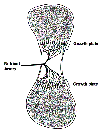
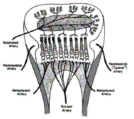
The second method is called endochondral ossification. This method describes the formation of most bones in the body. It begins with the precursory formation of a cartilage model covered with a perichondrium. Multiple blood vessels penetrate the cartilage to initiate bone formation and growth. The first blood vessel penetrates about mid-shaft and sends vessels to both ends of the cartilage model (Figure 6). This will set up the primary center of ossification. In addition, the vessel penetration causes osteoprogenitor cells to differentiate into osteoblasts and begin forming the periosteal collar and eventually the cortex of the diaphysis. The central regions form spongy trabecular patterns and are populated with red marrow. Second, the epiphyseal vessels penetrate the ends of the cartilage enlage and will form the secondary ossification centers. The vessels help arrange an environment around the growth plate that allows longitudinal growth. (Figure 7)
Figure 6

Figure 7


